Abstract
Introduction: Left ventricular hypertrophy is predictive of mortality in haemodialysis patients and occupies a very high proportion of cardiovascular complications. The aim of this study was to determine the prevalence of left ventricular hypertrophy in chronic renal failure patients under going hemodialysis at the CNHD. Materials and methods: This is a descriptive cross-sectional study in the Donka National Hemodialysis Center, running from February 01, 2024 to May 31, 2024. Chronic hemodialysis patients with electrical or echographic LVH were included in the study. Results: The number of chronic hemodialysis patients during the period was 405, among whom left ventricular hypertrophy was found in 140 patients or 34.57%. Chronic hemodialysis patients presenting with LVH during the study period had a mean age equal to 42.1±14.3 years, with a sex ratio= 2.3. Of 188 cardiac echograms performed, 112 cases (80%) had concentric hypertrophy and 28 cases (20%) had eccentric hypertrophy. Conclusion: The prevalence of left ventricular hypertrophy in chronic hemodialysis patients was found to be 34.57%. Large-scale studies in this chronic hemodialysis population are needed to investigate factors associated with left ventricular hypertrophy, in order to reduce cardiovascular morbidity and mortality.
|
Published in
|
Cardiology and Cardiovascular Research (Volume 9, Issue 3)
|
|
DOI
|
10.11648/j.ccr.20250903.11
|
|
Page(s)
|
88-94 |
|
Creative Commons
|

This is an Open Access article, distributed under the terms of the Creative Commons Attribution 4.0 International License (http://creativecommons.org/licenses/by/4.0/), which permits unrestricted use, distribution and reproduction in any medium or format, provided the original work is properly cited.
|
|
Copyright
|
Copyright © The Author(s), 2025. Published by Science Publishing Group
|
Keywords
Left Ventricular Hypertrophy, Chronic Hemodialysis Patients, Donka
1. Introduction
Left ventricular hypertrophy (LVH) corresponds to an increase in left ventricular mass due to an increase in myocyte size, often accompanied by myocardial fibrosis
| [1] | Mbaye A, Dodo B, Ngaïde AA, Sy NF, Babaka K, Mingou JS, et al. Left ventricular hypertrophy in black African subjects with artery hypertension. Ann Cardiol Angeiol (Paris) 2017; 66(4): 210-16. https://doi.org/ 10.1016/j.ancard.2017.04.011 |
[1]
.
It is characterized by a phenomenon of myocardial adaptation and an increase in cardiac afterload, requiring greater contraction force to eject the same volume. It is a predictive factor for mortality in chronic hemodialysis patients, and accounts for a very high proportion of cardiovascular complications
| [2] | Vigan J, Ahoui S, Hounsou D, Goudoté ACK, Vehounkpe Sacca J., Left ventricular hypertrophy in chronic hemodialysis patients at the CNHU-HKM in Cotonou. Nephrology Therapeutics NEPHRO. 2017; 14: 29-34. https://doi.org/10.1016/J.NEPHRO.2017.06.001 |
[2]
.
Multiple factors associated with declining renal function increase the risk of developing LVH, including anemia, hypertension, hypervolemia and disorders of mineral metabolism
. However, several other "non-traditional" risk factors have been identified in recent years that also contribute to the increased prevalence of CVD in this population. The most studied and influential factors in terms of cardiovascular risk are inflammation and oxidative stress
.
The links are close between renal and cardiac pathophysiology, and involve traditional cardiovascular risk factors, but also non-traditional risk factors linked to identified chronic renal failure
| [5] | Bayes-Genis A, De Antonio M, Vila J, Penafiel J, Galán A, Barallat J, et al. Head-to-Head Comparison of 2 Myocardial Fibrosis Biomarkers for Long-Term Heart Failure Risk Stratification. J Am Coll Cardiol 2014; 63: 158-66. https://doi.org/10.1016/j.cardfail.2015.07.017 |
[5]
. Among these specific factors are those directly linked to uraemia, such as hydrosodium overload, anaemia and hyperparathyroidism. Fluid retention contributes to arterial hypertension, LVH and increased arterial wall thickness. All these factors contribute to an increased cardiovascular risk. Patients suffering from chronic kidney disease (CKD) have a high rate of cardiovascular disease (CVD). Indeed, the prevalence of CVD is 10 to 30 times higher in these patients than in the general population
| [6] | Cissé MM, Tall LA, Faye M, Fall K, Faye Moustapha, Hadji KE, Diouf B et al. Evaluation of cardiac complications in Dakar chronic hemodialysis patients. Pan Afr Med J 2016.23.43.7227. https://doi.org/10.11604/pamj.2016.23.43.7227 |
[6]
.
Among cardiovascular complications, left ventricular hypertrophy accounts for a very high proportion of end-stage renal disease patients
| [7] | Zoccali C. Left Ventricular Mass Index as an Outcome Measure in Clinical Trials in Dialysis Patients: A Word of Caution. Am J Nephrol 2011; 33: 370-2. https://doi.org/10.1159/000324694 |
[7]
.
Several data are available in Asia, the United States and Europe on this subject,
In the United States Mc Cullough et al (2016) found a 75% prevalence of LVH in dialysis patients
| [8] | Mc Cullough PA, Chan CT, Weinhandl ED, Burkart JM, Bakris GL. Intensive Hemodialysis, Left Ventricular Hypertrophy, and Cardiovascular Disease. Am J Kidney Dis 2016; 68: S5-14. https://doi.org/10.1053/j.ajkd.2016.05.023 |
[8]
.
In Italy (2020), Nardi et al. showed a prevalence of LVH among hemodialysis patients in 62.8% of cases
| [9] | Nardi E, Mulè G, Giammanco A, Mattina A, Geraci G, Nardi C, et al. Left ventricular hypertrophy in chronic kidney disease: A diagnostic criteria comparison. Nutr Metab Cardiovasc Dis 2021; 31: 137-44. https://doi.org/10.1016/j.numecd.2020.08.028 |
[9]
.
In Japan, (2018) Kosaku et al. reported a 23.4% rate of LVH in hemodialysis patients
| [10] | Nitta K, Iimuro S, Imai E, Matsuo S, Makino H, Akizawa T, et al. Risk factors for increased left ventricular hypertrophy in patients with chronic kidney disease: findings from the CKD-JAC study. Clin Exp Nephrol 2019; 23: 85-98. https://doi.org/10.1007/s10157-018-1605-z |
[10]
.
In Algeria (2018), Kara et al. reported an LVH prevalence of 59.8% in their study
| [11] | Kara l, A. Abbou, R. Grari 1, D. Regagba, M. Ben Mansour et al. Left ventricular hypertrophy during chronic renal failure: prevalence and risk factors. Abstracts / Nephrology & Therapeutics. 2018; 14: 335-402. https://doi.org/10.1016/j.nephro.2018.07.198 |
[11]
.
In Morocco (2014), Eziani et al. had reported in a study carried out on 50 patients including 32 women and 18 men, 5 patients were hypertensive and anemia was found in 48% of cases
| [12] | Eziani M, Najdi A, Mikou S, Elhassani A, Akrichi MA, Hanin H, Mohamed A et al. Echocardiographic abnormalities in chronic hemodialysis patients: prevalence and risk factors. Pan Afr Med J 2014.18.216.4438. https://doi.org/10.11604/pamj.2014.18.216.4438 |
[12]
.
In Benin (2017), Vigan et al. reported a 57.5% prevalence of LVH
| [2] | Vigan J, Ahoui S, Hounsou D, Goudoté ACK, Vehounkpe Sacca J., Left ventricular hypertrophy in chronic hemodialysis patients at the CNHU-HKM in Cotonou. Nephrology Therapeutics NEPHRO. 2017; 14: 29-34. https://doi.org/10.1016/J.NEPHRO.2017.06.001 |
[2]
.
In Senegal (2016) Cissé Mouhamadou Moustapha et al. showed a 71.05% prevalence of LVH in hemodialysis patients
| [6] | Cissé MM, Tall LA, Faye M, Fall K, Faye Moustapha, Hadji KE, Diouf B et al. Evaluation of cardiac complications in Dakar chronic hemodialysis patients. Pan Afr Med J 2016.23.43.7227. https://doi.org/10.11604/pamj.2016.23.43.7227 |
[6]
.
In Guinea, a study by Balde Elhadj yaya et al. reported a prevalence of LVH of 85.7% in hypertensive patients
.
Management of hemodialysis patients has improved markedly, but with an increase in cardiovascular complications, which are the main cause of morbidity and mortality. The aim of this study was to determine the prevalence of left ventricular hypertrophy in chronic renal failure patients undergoing hemodialysis at the CNHD.
2. Materials and Methods
This is a descriptive cross-sectional study in the Centre National d'hémodialysis Donka, located within the CHU de Donka. This public dialysis center currently has 30 dialysis machines, and is also the only national center for public dialysis, renal disease referral and treatment of chronic end-stage renal failure.
Chronic hemodialysis patients constituted the study material, the study media were the medical records of hemodialysis patients, dialysis diaries, a Schiller brand Cardiovit FT-1 electrocardiograph, reports of cardiac echography performed free of charge at the CEMECO clinic by a cardiology specialist in search of ventricular hypertrophy and a survey form for data collection.
This is a descriptive cross-sectional study lasting four months, from February 01, 2024 to May 31, 2024.
Chronic hemodialysis patients were targeted during the study period; the study population consisted of hemodialysis patients with a clinical presentation of right, left or congestive heart failure and cardiovascular risk factors despite regularly monitored hemodialysis. Only chronic hemodialysis patients with LVH on cardiac ultrasound were included in the study. ECG was not performed in patients without informed consent to orient the diagnosis of LVH, prior to confirmatory cardiac ultrasound.
Recruitment involved all chronic hemodialysis patients during the study period who met the inclusion criteria. A minimum sample of 140 patients with LVH on cardiac ultrasound was obtained; data were collected on an individual survey form.
Variables were defined by epidemiological data (frequency, age, sex), clinical data (initial nephropathy, duration of dialysis), ECG and cardiac ultrasound data.
2.1. Epidemiological Data
1) Prevalence: Corresponds to the proportion of hemodialysis patients with left ventricular hypertrophy over a study period.
2) Age: Divided into 10-year age brackets, to determine average age.
3) Gender: to determine the M/F sex ratio.
2.2. Causal Nephropathy
Among the causes we have:
1. Chronic glomerular nephropathy
It was characterized by:
(1) A history of recurrent edema of the lower limbs;
(2) Proteinuria ≥1.5 g/24h;
(3) High blood pressure.
2. Chronic interstitial nephropathy
It was characterized by:
(1) A history of recurrent urinary tract infection or urinary lithiasis;
(2) No proteinuria or moderate proteinuria<1 g/24h;
(3) Germ-free leukocyturia.
3. Vascular nephropathy
Clinical and biological evidence in favour:
(1) A history of arterial hypertension;
(2) Moderate proteinuria ≤ 1 g/l;
(3) Kir Kendall stage 2 or 3 hypertensive retinopathy;
(4) Concentric LVH.
4. Diabetic nephropathy
The patient had had diabetes for several years with:
(1) Micro-albuminuria˃ 30 mg/24h or macro-albuminuria (proteinuria) >300 mg/24h;
(2) Diabetic retinopathy on fundus;
5. Indeterminate nephropathies: all nephropathies that have not been classified in one of the nosological groups.
6. Hemodialysis life time: the time spent on hemodialysis since the date of initiation of hemodialysis, measured in months or years.
2.2.1. ECG Data
the Sokolow-Lyon index and the Cornell index (used for hypertension) for diagnosing LVH. Thus, we considered LVH in front of the index of:
1) Sokolow Lyon: If the sum of SV1+(RV5 or RV6) is ˃35 mm for patients aged 40 or over.
2) Cornell: If the sum of RVL+SV3 amplitudes ˃ 20 mm in women and ˃28 mm in men. ECG results were interpreted under the assistance of a cardiologist.
2.2.2. Cardiac Ultrasound
To detect hypertrophy and/or dilation of the heart chambers, heart failure, pericarditis or valvulopathy. Cardiac ultrasound is performed free of charge at the CEMECO clinic by a doctor specializing in cardiology.
1) Ultrasound LVH: When the thicknesses of the interventricular septum and the posterior wall of the left ventricle are greater than 11 mm at the end of diastole.
2) Concentric LVH was defined as a left ventricular mass index greater than 115 g/m2 in men and greater than 95 g/m2 in women, with an RWT greater than 0.42.
3) Eccentric LVH was defined as a left ventricular mass index greater than 115 g/m2 in men and greater than 95 g/m2 in women, with an RWT less than 0.42.
2.3. Data Were Collected on Survey Forms
A data base was created using the Kobocollect application for data entry, then exported to SPSS (Statistical Package for Social Science) version 2.1.0 for statistical analysis. Qualitative variables are interpreted in terms of frequencies and proportions, while quantitative variables are interpreted in terms of averages.
3. Results
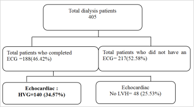
The number of chronic hemodialysis patients during the period was 405, of whom 188 had completed electrocardiograms. Left ventricular hypertrophy was found on cardiac echography in 140 patients, or 34.57% (
Figure 1).
Figure 1. Frequency of chronic haemodialysis patients with left ventricular hypertrophy during the study period at the Donka national haemodialysis center.
Chronic haemodialysis patients presenting with LVH during the study period had a mean age equal to 42.1± 14.3 years, with extremes of 13 and 75 years; the 36-50 age group accounted for 77 or 55% (
Table 1).
Table 1. Age distribution of chronic hemodialysis patients with left ventricular hypertrophy during the study period.
Age ranges | Number (N=140) | Percentage |
≤ 20 years | 4 | 2,9 |
21 to 35 years old | 32 | 22,8 |
36 to 50 years | 77 | 55 |
51 to 65 years | 22 | 15,7 |
˃ 65 years | 5 | 3,6 |
Total | 140 | 100 |
Mean age: 42.1±14.3 years
Extremes: 13 and 75 years
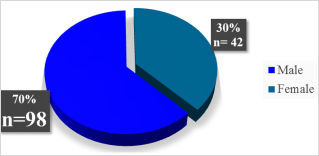
Of 140 chronic hemodialysis patients with LVH, 98 (70%) were predominantly male, with a sex ratio of 2.3 (
Figure 2).
Figure 2. Distribution of chronic haemodialysis patients with left ventricular hypertrophy during the study period by gender.
Of 140 patients under going chronic hemodialysis during the study period, the nephropathy responsible for end-stage renal failure was dominated by vascular nephropathy in 53 patients (37.86%) (
table 2).
Table 2. Distribution of chronic hemodialysis patients with left ventricular hypertrophy during the study period, according to causal nephropathies.
Initial kidney disease | Number (N=140) | % |
Glomerular nephropathy | 47 | 33,57 |
Nephropathy indeterminate | 24 | 17,14 |
Vascular nephropathy | 53 | 37,86 |
Diabetic nephropathy | 13 | 9,29 |
Chronic tubulointerstitial nephropathy | 3 | 2,14 |
Out of 140 chronic hemodialysis patients, 56 (40%) were in the 3-9 month range (
Table 3).
Table 3. Distribution of chronic haemodialysis patients with left ventricular hypertrophy during the study period, according to length of haemodialysis.
Length of time on hemodialysis (in months) | Numbers (N=140) | Percentages |
3-9 | 56 | 40 |
10-16 | 13 | 9,29 |
17-23 | 24 | 17,14 |
>24 | 48 | 34,28 |
Out of a total of 188 ECG cases, 145 cases or 77.13% showed left ventricular hypertrophy according to the Sokolow index with a mean =44.9±16.7 mm. According to the average Cornell index= 23.5±11.2 mm, with a predominance in men with an index≥ 28 mm in 38 cases or 20.2%, and in women with an index ≥20 mm in 38 cases or 20.2% (
Table 4).
Table 4. Distribution of chronic hemodialysis patients with left ventricular hypertrophy during the study period, by ECG result.
ECG | Numbers (N = 188) | Percentages |
Yes | 188 | 100 |
Sokolow-Lyon index (mm) |
Normal (< 35) | 43 | 22,87 |
≥ 35 | 145 | 77,13 |
Cornell index (mm) |
< 20 (Female) | 34 | 18,1 |
< 28 (male) | 78 | 41,5 |
≥ 20 (Female) | 38 | 20,2 |
≥ 28 (Male) | 38 | 20,2 |
Mean Sokolow index: 44.9 ±16.7 mm Extremes: 6 and 97.2 mm
Average Cornell index: 23.5 ±11.2 mm Extremes: 2.6 and 70.8 mm
Of 140 cardiac ultrasounds performed, 112 cases (80%) had concentric hypertrophy and 28 cases (20%) had eccentric hypertrophy (
Table 5).
Table 5. Distribution of chronic hemodialysis patients with left ventricular hypertrophy during the study period, according to cardiac ultrasound findings.
Cardiac ultrasound results | Number (N=140) | Percentages |
Concentric HVG | 112 | 80 |
HVG eccentric | 28 | 20 |
The association between gender and left ventricular hypertrophy on cardiac ultrasound yielded a statistically significant result with P-value =0.003; more specifically, male gender had a positive influence on left ventricular hypertrophy (
Table 6).
Table 6. Distribution of chronic hemodialysis patients with left ventricular hypertrophy on cardiac echocardiography and associated gender.
HVG | HVG Concentric | HVG Eccentric | Total | Chi-2 Square | P-Value |
Gender | F | 33 (23,57%) | 7 (5%) | 40 | 138,526 | 0,4 |
M | 78 (55,71%) | 20 (14,29%) | 98 | 0,003 |
Total | | 111 (79,29%) | 27 (19,29%) | 138 | 140 | |
4. Discussion
The present study was conducted at the Donka CNHD. We enrolled 140 patients over a 4-month period, all of whom met the inclusion criteria.
The prevalence of LVH reported in our series was 34.57% on cardiac ultrasound. The mean age was 42.1 ±13.3 years. Men were the most affected in our series, accounting for 70%, i.e. a sex ratio of 2.3. The nephropathy responsible for end-stage renal failure was dominated by vascular nephropathy in 53 patients, i.e. 37.86%. Out of 140 chronic hemodialysis patients presenting with left ventricular hypertrophy on cardiac ultrasound, the age, expressed in months, of 56 patients (40%) was between 3 and 9 months. In our study, 140 patients showed left ventricular hypertrophy on cardiac ultrasound. LVH was concentric in 112 patients (80%), and 28 patients had eccentric LVH (20%). The association is particularly significant, with P-value =0.003, between male sex and positive influence on left ventricular hypertrophy.
Several studies have been carried out on the subject, and each has found a greater or lesser frequency of LVH in dialysis patients.
Chargui et al. reported a higher prevalence than ours, with 65% LVH in a study of 60 patients carried out in 2021
| [14] | Soumaya Chargui, Emna Allouche, Mariem Hajji, Wiem Dkhil, Sahar Agrebi, Habib Ben Ahmed, Khaled Ezzaouia et al. Left ventricular hypertrophy in hemodialysis patients: prevalence, electrical, echographic study and risk factors. 2022; 18(4,): 247-254. https://doi.org/10.1016/j.nephro.2021.10.003 |
[14]
. Faye et al. reported a 50.57% prevalence of LVH in a study of 97 hemodialysis patients
| [15] | Maria Faye, Ahmed Tall Lemrabott, Bacary Ba, Moustapha Faye, Fatima Ezzahra Boudal, Mansour Mbengue et al. Electrical and Echocardiographic abnormalities in Chronic Hemodialysis Patients in Dakar. Health Sci. Dis: 2022; 23(6): 21-25. www.hsd-fmsb.org |
[15]
. Foley et al. found 39.4% concentric LVH in their patients
| [16] | Foley RN, Parfrey PS, Harnett JD, Kent GM, Murray DC, Barre PE. Impact of hypertension on cardiomyopathy, morbidity and mortality in end-stage renal disease. Kidney Int 1996; 49: 1379-85. https://doi.org/10.1038/ki.1996.194 |
[16]
. A study by Levin et al. reported that concentric LVH appears very early in the natural history of chronic renal failure
| [17] | Levin A, Singer J, Thompson CR, Ross H, Lewis M. Prevalent left ventricular hypertrophy in the predialysis population: identifying opportunities for intervention. Am J Kidney Dis 1996; 27: 347-54. https://doi.org/10.1016/s0272-6386(96)90357-1 |
[17]
. Zhou et al., on the other hand, found 71.8% eccentric LVH
| [18] | Zhou YL, Liu J, Ma L, Sun F, Shen Y, Huang J, et al. Impact of dry weight determined by calf bioimpedance ratio on carotid stiffness and left ventricular hypertrophy in haemodialysis patients. Artificial Organs. 2014; 38: 327-34. https://doi.org/10.1111/aor.12156 |
[18]
.
Ashok Vankayala et al. obtained a mean age of 43.27±10.1, higher than our results
| [19] | Ashok Vankayala, Kamal Lochan Behera, DSSK Raju, Suresh Babu Sayana. A study of left ventricular hypertrophy in patients with stage three to five chronic kidney disease. Int J Res Med Sci. 2019; 7(5): 1511-1514. https://doi.org/10.18203/2320-6012.ijrms20191502 |
[19]
. Vigan Jacques et al. reported a lower result, with a male predominance of 61% and a sex ratio of 1.6
| [2] | Vigan J, Ahoui S, Hounsou D, Goudoté ACK, Vehounkpe Sacca J., Left ventricular hypertrophy in chronic hemodialysis patients at the CNHU-HKM in Cotonou. Nephrology Therapeutics NEPHRO. 2017; 14: 29-34. https://doi.org/10.1016/J.NEPHRO.2017.06.001 |
[2]
. The difference in study population, study duration, sample size, collection method, socio-economic factors, despite the same LVH assessment criteria, could be the reasons for this difference.
The study's limitations in comparison with the literature are mainly due to the type of study, which is cross-sectional in this series, but also to the difference in study duration. However, the study populations are the same. The results of these different studies are super imposable, giving the study internal and external validity. This comparison gives a generalizability to the results obtained in relation to certain data in the current literature.
1) Ethical considerations: in the field, informed consent was obtained from participants before submitting them to the questionnaire, and strict confidentiality was observed.
2) Study limitations: patients with central venous catheters and those with fistulas were reluctant to have electrodes placed, and the lack of informed consent corresponding to 217 hemodialysis patients (52.58%) despite information on non-invasive cardiac ultrasound.
3) Conclusion: patients suffering from chronic renal failure have a high rate of cardiovascular disease. The prevalence of cardiovascular disease is 10 to 30 times higher in these patients than in the general population. In this study, the prevalence of left ventricular hypertrophy in chronic hemodialysis patients was found to be 34.57%. The mean age of these patients was 42.1±14 years, with a male predominance. Cardiac ultrasonography found 112 cases of concentric ventricular hypertrophy in 80% of chronic hemodialysis patients. Large-scale studies in this chronic hemodialysis population are needed to investigate factors associated with this left ventricular hypertrophy, in order to reduce cardiovascular morbidity and mortality.
Abbreviations
CHU | Centre Hospitalo-Universitaire |
CNHD | Donka National Hemodialysis Center |
ECG | Electrocardiogram |
LVH | Left Ventricular Hypertrophy |
BMI | Body Mass Index |
CKD | Chronic Kidney Disease |
CVD | Cardiovascular Disease |
Acknowledgments
To my teachers, Professor KABA Mohamed Lamine and Professor Bah Alpha Oumar, your love of a job well done and your skills have encouraged me to pur sue research. Please accept the expression of my deepest gratitude. Dr Bangoura Soriba, Dr Traoré Aly, thank you for your commitment and perseverance from the protocol to submission of the manuscript, and allow me to thank you for your scientific rigor. Thank you to the readers of this journal who provided critical reading of the manuscript to improve the scientific continuum. Please accept my deepest gratitude.
Conflicts of Interest
The authors declare no conflicts of interest.
References
| [1] |
Mbaye A, Dodo B, Ngaïde AA, Sy NF, Babaka K, Mingou JS, et al. Left ventricular hypertrophy in black African subjects with artery hypertension. Ann Cardiol Angeiol (Paris) 2017; 66(4): 210-16.
https://doi.org/ 10.1016/j.ancard.2017.04.011
|
| [2] |
Vigan J, Ahoui S, Hounsou D, Goudoté ACK, Vehounkpe Sacca J., Left ventricular hypertrophy in chronic hemodialysis patients at the CNHU-HKM in Cotonou. Nephrology Therapeutics NEPHRO. 2017; 14: 29-34.
https://doi.org/10.1016/J.NEPHRO.2017.06.001
|
| [3] |
Wanner C, Amann K, Shoji T. The heart and vascular system in dialysis. The Lancet 2016; 388: 276-84.
https://doi.org/10.1016/S0140-6736(16)30508-6
|
| [4] |
Madore F. Vascular risk factors and renal failure. Med Sci 2004: 1100-3.
https://doi.org/10.1051/medsci/200420121100
|
| [5] |
Bayes-Genis A, De Antonio M, Vila J, Penafiel J, Galán A, Barallat J, et al. Head-to-Head Comparison of 2 Myocardial Fibrosis Biomarkers for Long-Term Heart Failure Risk Stratification. J Am Coll Cardiol 2014; 63: 158-66.
https://doi.org/10.1016/j.cardfail.2015.07.017
|
| [6] |
Cissé MM, Tall LA, Faye M, Fall K, Faye Moustapha, Hadji KE, Diouf B et al. Evaluation of cardiac complications in Dakar chronic hemodialysis patients. Pan Afr Med J 2016.23.43.7227.
https://doi.org/10.11604/pamj.2016.23.43.7227
|
| [7] |
Zoccali C. Left Ventricular Mass Index as an Outcome Measure in Clinical Trials in Dialysis Patients: A Word of Caution. Am J Nephrol 2011; 33: 370-2.
https://doi.org/10.1159/000324694
|
| [8] |
Mc Cullough PA, Chan CT, Weinhandl ED, Burkart JM, Bakris GL. Intensive Hemodialysis, Left Ventricular Hypertrophy, and Cardiovascular Disease. Am J Kidney Dis 2016; 68: S5-14.
https://doi.org/10.1053/j.ajkd.2016.05.023
|
| [9] |
Nardi E, Mulè G, Giammanco A, Mattina A, Geraci G, Nardi C, et al. Left ventricular hypertrophy in chronic kidney disease: A diagnostic criteria comparison. Nutr Metab Cardiovasc Dis 2021; 31: 137-44.
https://doi.org/10.1016/j.numecd.2020.08.028
|
| [10] |
Nitta K, Iimuro S, Imai E, Matsuo S, Makino H, Akizawa T, et al. Risk factors for increased left ventricular hypertrophy in patients with chronic kidney disease: findings from the CKD-JAC study. Clin Exp Nephrol 2019; 23: 85-98.
https://doi.org/10.1007/s10157-018-1605-z
|
| [11] |
Kara l, A. Abbou, R. Grari 1, D. Regagba, M. Ben Mansour et al. Left ventricular hypertrophy during chronic renal failure: prevalence and risk factors. Abstracts / Nephrology & Therapeutics. 2018; 14: 335-402.
https://doi.org/10.1016/j.nephro.2018.07.198
|
| [12] |
Eziani M, Najdi A, Mikou S, Elhassani A, Akrichi MA, Hanin H, Mohamed A et al. Echocardiographic abnormalities in chronic hemodialysis patients: prevalence and risk factors. Pan Afr Med J 2014.18.216.4438.
https://doi.org/10.11604/pamj.2014.18.216.4438
|
| [13] |
Balde E, Barry I, Balde M, Beavogui M, Diop I, Jouven X, Toure Set. Definitive cardiac pacing: first experience in Guinea. Tropical-cardiology 2013; 06: 44-55.
https://tropical-cardiology.com/Accueil/index.php/2013-08-10-06-44-55
|
| [14] |
Soumaya Chargui, Emna Allouche, Mariem Hajji, Wiem Dkhil, Sahar Agrebi, Habib Ben Ahmed, Khaled Ezzaouia et al. Left ventricular hypertrophy in hemodialysis patients: prevalence, electrical, echographic study and risk factors. 2022; 18(4,): 247-254.
https://doi.org/10.1016/j.nephro.2021.10.003
|
| [15] |
Maria Faye, Ahmed Tall Lemrabott, Bacary Ba, Moustapha Faye, Fatima Ezzahra Boudal, Mansour Mbengue et al. Electrical and Echocardiographic abnormalities in Chronic Hemodialysis Patients in Dakar. Health Sci. Dis: 2022; 23(6): 21-25.
www.hsd-fmsb.org
|
| [16] |
Foley RN, Parfrey PS, Harnett JD, Kent GM, Murray DC, Barre PE. Impact of hypertension on cardiomyopathy, morbidity and mortality in end-stage renal disease. Kidney Int 1996; 49: 1379-85.
https://doi.org/10.1038/ki.1996.194
|
| [17] |
Levin A, Singer J, Thompson CR, Ross H, Lewis M. Prevalent left ventricular hypertrophy in the predialysis population: identifying opportunities for intervention. Am J Kidney Dis 1996; 27: 347-54.
https://doi.org/10.1016/s0272-6386(96)90357-1
|
| [18] |
Zhou YL, Liu J, Ma L, Sun F, Shen Y, Huang J, et al. Impact of dry weight determined by calf bioimpedance ratio on carotid stiffness and left ventricular hypertrophy in haemodialysis patients. Artificial Organs. 2014; 38: 327-34.
https://doi.org/10.1111/aor.12156
|
| [19] |
Ashok Vankayala, Kamal Lochan Behera, DSSK Raju, Suresh Babu Sayana. A study of left ventricular hypertrophy in patients with stage three to five chronic kidney disease. Int J Res Med Sci. 2019; 7(5): 1511-1514.
https://doi.org/10.18203/2320-6012.ijrms20191502
|
Cite This Article
-
APA Style
Tégui, C. M. L., Soriba, B., Djasrabe, N. S., Aly, T., Moussa, T., et al. (2025). Prevalence of Left Ventricular Hypertrophy in Chronic Renal Failure Patients Under Going Hemodialysis in Donka. Cardiology and Cardiovascular Research, 9(3), 88-94. https://doi.org/10.11648/j.ccr.20250903.11
 Copy
|
Copy
|
 Download
ACS Style
Tégui, C. M. L.; Soriba, B.; Djasrabe, N. S.; Aly, T.; Moussa, T., et al. Prevalence of Left Ventricular Hypertrophy in Chronic Renal Failure Patients Under Going Hemodialysis in Donka. Cardiol. Cardiovasc. Res. 2025, 9(3), 88-94. doi: 10.11648/j.ccr.20250903.11
Copy
|
Download
AMA Style
Tégui CML, Soriba B, Djasrabe NS, Aly T, Moussa T, et al. Prevalence of Left Ventricular Hypertrophy in Chronic Renal Failure Patients Under Going Hemodialysis in Donka. Cardiol Cardiovasc Res. 2025;9(3):88-94. doi: 10.11648/j.ccr.20250903.11
Copy
|
Download
-
@article{10.11648/j.ccr.20250903.11,
  author = {Camara Mohamed Lamine Tégui and Bangoura Soriba and Nguena Shallom Djasrabe and Traore Aly and Traore Moussa and Tolno Abou and Diakite Fousseny and Kaba Mohamed Lamine},
  title = {Prevalence of Left Ventricular Hypertrophy in Chronic Renal Failure Patients Under Going Hemodialysis in Donka
},
  journal = {Cardiology and Cardiovascular Research},
  volume = {9},
  number = {3},
  pages = {88-94},
  doi = {10.11648/j.ccr.20250903.11},
  url = {https://doi.org/10.11648/j.ccr.20250903.11},
  eprint = {https://article.sciencepublishinggroup.com/pdf/10.11648.j.ccr.20250903.11},
  abstract = {Introduction: Left ventricular hypertrophy is predictive of mortality in haemodialysis patients and occupies a very high proportion of cardiovascular complications. The aim of this study was to determine the prevalence of left ventricular hypertrophy in chronic renal failure patients under going hemodialysis at the CNHD. Materials and methods: This is a descriptive cross-sectional study in the Donka National Hemodialysis Center, running from February 01, 2024 to May 31, 2024. Chronic hemodialysis patients with electrical or echographic LVH were included in the study. Results: The number of chronic hemodialysis patients during the period was 405, among whom left ventricular hypertrophy was found in 140 patients or 34.57%. Chronic hemodialysis patients presenting with LVH during the study period had a mean age equal to 42.1±14.3 years, with a sex ratio= 2.3. Of 188 cardiac echograms performed, 112 cases (80%) had concentric hypertrophy and 28 cases (20%) had eccentric hypertrophy. Conclusion: The prevalence of left ventricular hypertrophy in chronic hemodialysis patients was found to be 34.57%. Large-scale studies in this chronic hemodialysis population are needed to investigate factors associated with left ventricular hypertrophy, in order to reduce cardiovascular morbidity and mortality.
},
 year = {2025}
}
Copy
|
Download
-
TY  - JOUR
T1  - Prevalence of Left Ventricular Hypertrophy in Chronic Renal Failure Patients Under Going Hemodialysis in Donka

AU  - Camara Mohamed Lamine Tégui
AU  - Bangoura Soriba
AU  - Nguena Shallom Djasrabe
AU  - Traore Aly
AU  - Traore Moussa
AU  - Tolno Abou
AU  - Diakite Fousseny
AU  - Kaba Mohamed Lamine
Y1  - 2025/07/09
PY  - 2025
N1  - https://doi.org/10.11648/j.ccr.20250903.11
DO  - 10.11648/j.ccr.20250903.11
T2  - Cardiology and Cardiovascular Research
JF  - Cardiology and Cardiovascular Research
JO  - Cardiology and Cardiovascular Research
SP  - 88
EP  - 94
PB  - Science Publishing Group
SN  - 2578-8914
UR  - https://doi.org/10.11648/j.ccr.20250903.11
AB  - Introduction: Left ventricular hypertrophy is predictive of mortality in haemodialysis patients and occupies a very high proportion of cardiovascular complications. The aim of this study was to determine the prevalence of left ventricular hypertrophy in chronic renal failure patients under going hemodialysis at the CNHD. Materials and methods: This is a descriptive cross-sectional study in the Donka National Hemodialysis Center, running from February 01, 2024 to May 31, 2024. Chronic hemodialysis patients with electrical or echographic LVH were included in the study. Results: The number of chronic hemodialysis patients during the period was 405, among whom left ventricular hypertrophy was found in 140 patients or 34.57%. Chronic hemodialysis patients presenting with LVH during the study period had a mean age equal to 42.1±14.3 years, with a sex ratio= 2.3. Of 188 cardiac echograms performed, 112 cases (80%) had concentric hypertrophy and 28 cases (20%) had eccentric hypertrophy. Conclusion: The prevalence of left ventricular hypertrophy in chronic hemodialysis patients was found to be 34.57%. Large-scale studies in this chronic hemodialysis population are needed to investigate factors associated with left ventricular hypertrophy, in order to reduce cardiovascular morbidity and mortality.

VL  - 9
IS  - 3
ER  -
Copy
|
Download